
Mastering the Wound Healing Cascade: From Biological Phases to Bedside Care
Understanding the mechanics of tissue repair is fundamental to advanced wound management and chronic wound care across care settings nationwide. While standard textbooks often outline the wound healing cascade in neat, isolated stages, clinical practice tells a different story. Healing is a fluid, continuous process where each phase overlaps and heavily influences the success of the next.
This clinical framework is highlighted in the video“Micro-session: The Wound Healing Cascade,”presented byWound Care University, an educational division of WoundCentrics. As the clinical education arm of WoundCentrics, Wound Care University provides evidence-based, accredited training and continuing education in hyperbaric medicine and advanced wound care for physicians, advanced practice providers, and allied health professionals throughout the United States.
When a wound fails to progress through this cascade, it stalls—often becoming a chronic wound that consumes valuable clinical resources and impacts patient quality of life. By mapping the cellular events of healing to practical interventions, healthcare providers can better identify local and systemic barriers, reset the microenvironment, and drive wounds toward closure in hospitals, outpatient clinics, and post-acute settings nationwide.
The Four Overlapping Phases of Healing
The wound healing process is commonly described in four overlapping phases that form a dynamic cascade rather than a simple linear sequence.
Hemostasis → Inflammation → Proliferation → Remodeling
(Immediate) → (Days 1–4) → (Days 4–21) → (21 Days – Months)
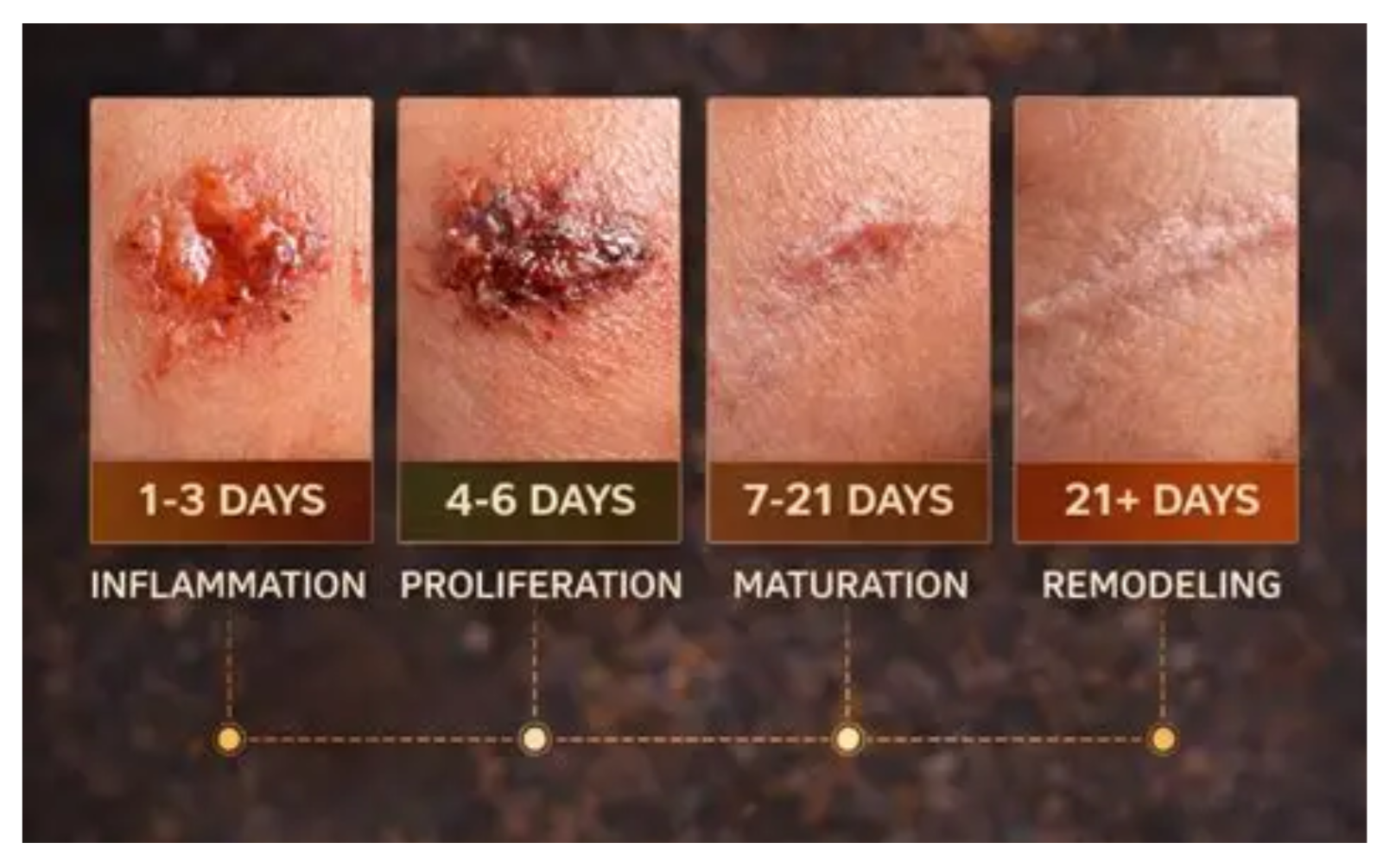
Each phase has distinct cellular players, molecular drivers, and bedside implications, yet they function as a coordinated system to restore tissue integrity.
Phase 1 – Hemostasis (Immediate Response)
Immediately following tissue injury, the body acts to prevent blood loss and establish a temporary matrix.
• Vascular constriction and platelet plug: Blood vessels constrict, and platelets aggregate upon contacting exposed extracellular collagen.
• Biological fibrin clot: Coagulation pathways activate, stabilizing the initial plug into a robust fibrin clot.
• Provisional scaffold: This clot acts as a biological reservoir—trapping repair cells and storing key growth factors such as PDGF (platelet-derived growth factor) and TGF-β (transforming growth factor-beta) to initiate downstream healing.
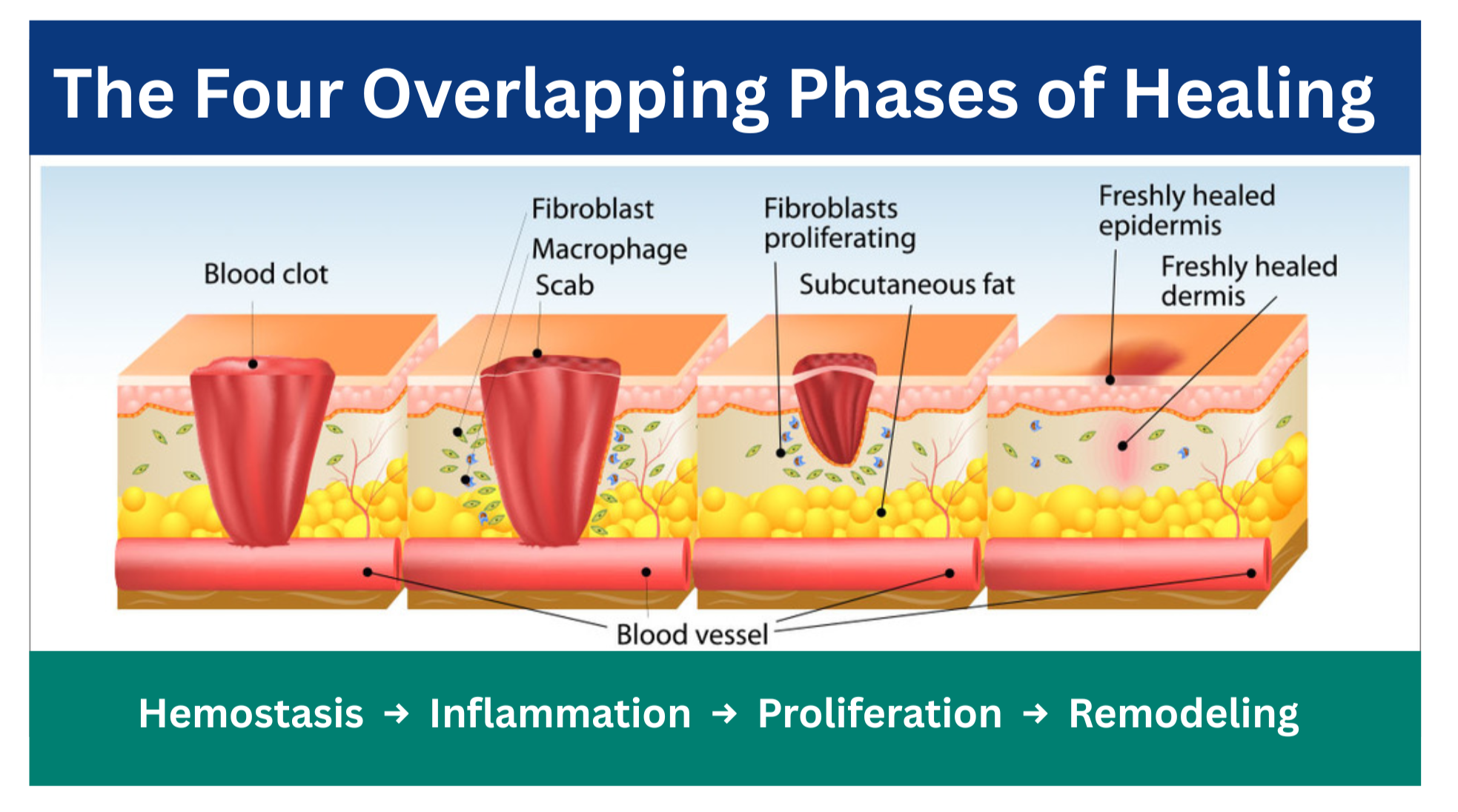
At the bedside, rapid hemostasis creates the foundation for subsequent debridement, infection prevention, and moisture management strategies used by wound care teams across the country.
Phase 2 – Inflammation (Cellular Clean-Up and Signaling)
As blood flow stabilizes, immune cells flood the wound bed to clear debris and prevent infection.
• Neutrophil invasion: Within hours, neutrophils phagocytose bacteria and devitalized tissue while releasing proteases and reactive oxygen species.
• Macrophage transition: Monocytes differentiate into M1-like (pro-inflammatory) macrophages to continue cleanup, then transition to M2-like (pro-repair) phenotypes that release growth factors to drive tissue reconstruction.
• Cytokine signals: Interleukins such as IL‑1 and IL‑6 and TNF‑α regulate vascular permeability and cell recruitment.
Clinical takeaway: Balance is critical. Under-inflammation increases infection risk, while persistent or dysregulated inflammation can trap a wound in a chronic, non-healing state—one of the most common challenges in advanced wound care nationally.
Phase 3 – Proliferation (Rebuilding Structure)
Once the wound bed is sanitized, structural rebuild begins.
Angiogenesis: Guided by VEGF (vascular endothelial growth factor), endothelial cells sprout new capillary networks, forming healthy, ruby-red granulation tissue.
Extracellular matrix deposition: Fibroblasts synthesize preliminary type III collagen, establishing early tensile strength.
Closure and contraction: Keratinocytes migrate from wound edges to re-epithelialize the surface, while myofibroblasts contract the wound margins.
During this phase, clinicians across the United States rely on evidence-based dressing selection, moisture balance, and offloading strategies to support granulation and protect fragile new tissue.
Phase 4 – Remodeling (Maturation and Refinement)
The final phase reshapes and strengthens newly formed tissue over months to years.
• Collagen transition: Type III collagen is systematically replaced by stronger type I collagen, which cross-links along lines of mechanical stress.
• Scar maturation: Excess blood vessels regress, causing hyperemic scars to gradually blanch.
• Tensile limits: Remodeled skin reaches a maximum of approximately 80% of its original tensile strength.
Nationwide, wound care teams emphasize scar management, long-term offloading, and recurrence prevention during this maturation phase.
Why Wounds Stall: Identifying Barriers to Healing
Chronic wounds frequently become “stuck” in the inflammatory phase. In this state, elevated levels of matrix metalloproteinases (MMPs) and proteases outpace their natural inhibitors (TIMPs), degrading extracellular matrix and essential growth factors before new tissue can build.
To restart the cascade, clinicians must identify and address both local and systemic barriers
Barrier category – Key factors and impediments
Local barriers: Biofilm burden, tissue ischemia, sustained pressure or shear, localized edema, or uncontrolled exudate.
Systemic barriers: Malnutrition (protein and micronutrient deficiencies), uncontrolled diabetes, peripheral vascular disease, and therapies such as immunosuppressants or steroids.
These challenges are common across U.S. inpatient, outpatient, and post-acute care environments, making standardized, cascade-based approaches especially valuable.
Translating Biology to Bedside Practice
Effective wound management relies on removing physiological roadblocks and matching orders to the patient’s current phase of healing.
1. Perform effective debridement: Remove bioburden, necrotic tissue, and senescent cells to clear MMP-heavy exudate and convert chronic tissue back into an acute healing state.
2. Optimize the microenvironment: Select advanced dressings that maintain balanced moisture, provide thermal stability, and safeguard delicate peri-wound skin.
3. Resolve underlying etiologies: Restore arterial perfusion where possible, apply compression for venous hypertension, and implement appropriate pressure offloading.
4. Support host systemic health: Optimize blood glucose and ensure adequate dietary protein intake to supply necessary amino acids for extracellular matrix synthesis.
Aligning orders with the wound’s current phase helps healthcare teams across the country standardize practice, protect new tissue growth, and improve outcomes in diverse care settings.
Learn More with WoundCentrics and Wound Care University
By aligning clinical treatments with the underlying biology of the wound healing cascade, healthcare teams can systematically eliminate barriers, protect new tissue growth, and improve patient outcomes nationwide. WoundCentrics partners with hospitals, health systems, and post-acute providers throughout the United States to deliver comprehensive advanced wound care and hyperbaric medicine programs.
To explore more educational videos and training opportunities, visit Wound Care University, the clinical education arm of WoundCentrics, or watch the full micro-session on YouTube: “Micro-session: The Wound Healing Cascade.”